Dr. Dipankar Das, D08987, Dr. Harsha Bhattacharjee, Dr. Satyen Deka
Full text AIOC 2017-FP 509:
Section: Neuro-ophthalmology
Title of the paper: Optic nerve anatomy for future biometric signature
Chief and presenting author-Dipankar Das (D08987)
Co-authors: Harsha Bhattacharjee, Satyen Deka
Sri Sankaradeva Nethralaya, Guwahati, Assam, INDIA
Synopsis
Biometric system works on the basis of recognition of patterns. There are various existing biometric system for identification such as fingerprinting, iris and retinal vessels pattern. We present the anatomical morphology of optic nerve transverse section as one of the additional unique biometric signature in human beings.
Methods: Observational case series in ocular pathology laboratory where (n=200) transverse optic nerve sections from enucleated eyes were studied. Enucleated specimens were received in laboratory for various indications and after written consent. They were documented in AxioSkop Zeiss microscope MRc camera. Special stains were done.
Results: The septas of optic nerve, nerve bundle and inter-septal vascularisations in the optic nerve were unique for each transverse section specimen at the point of cut, there was no situation where similarities were observed in the studied specimens.
Conclusion: Optic nerve can be an important biometric signature in humans.
Introduction:
As a unique characteristic, we have different biometric indicators for each and every human being.1,2,3,4,5 Finger printing, poroscopy, iris and retinal vessels scans are already there and are established trait for unique identification.1 Some of the biometric indicators such as fingerprint or iris color and configuration can be modifiable.2,3 Optic nerve, as a cranial nerve is unique. It is only cranial nerve that comes out of the cranium and only nerve to be visualized directly by ophthalmoscope. We have seen a characteristic pattern of anatomical arrangement of transected optic nerve along with the micro-vessels in the septas separating the bundles of optic nerve, which could be a unique anatomical characteristic at the point of transverse slice. No two optic nerves has same configuration in the study.
Objectives: To present the findings of anatomical and morphological optic nerve transections as human biometric signatures.
Methodology: This study was approved by institutional research and ethics committee.
- Place of study: Sri Sankaradeva Nethralaya, Guwahati, Assam, India.
- Type of study: Laboratory based observational study
- Material & methods: A total of (n=200) transected optic nerve sections were studied. All the eyeball specimens were enucleated for various indications and after proper written consent. Normal hematoxylin eosin (H&E) sections were studied for mapping the optic nerves. Optic nerves were observed for the following patterns: 1. Distribution of septas and 2. The blood vessels within the septas. [Figure 1,2]
Some of the optic nerve sections were seen in unstained sections under the polarizer [Figure 3]. Special stains such as Masson’s trichrome was done to see the collagen septas containing the nerve bundles. The mapping of optic nerve was documented in diffraction photography, using polarizer and different objective of ZEISS Axioskop 40 with AxioCam MRc camera. Some of these images were rendered in gray scale and the white images on the dark background were adjusted to enhance the contrast. The images were analyzed uniformly based on magnifications.
Results: The collagen septas of the optic nerve containing the bundles of nerves had unique distributions and their orientations at the point of transverse cut of the optic nerves were exclusive. No two septal distributions in optic nerve had similar pattern. The micro blood vessels within the collagen septas had a characteristic position distribution which did not match with any other optic nerve sections.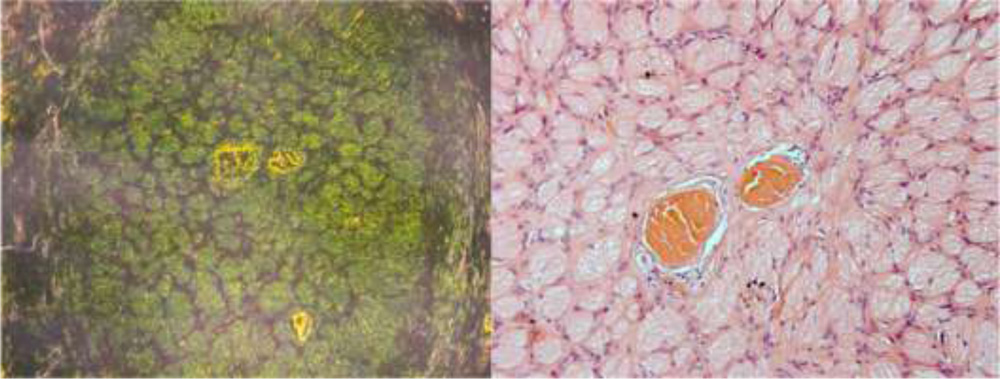
Figure 1: Transverse slice of optic nerve in diffraction and H&E Stain (20x, 40 x)
Figure 2: H&E and Masson trichrome stain with septas and inter-septal blood vessels (20x, 40 x)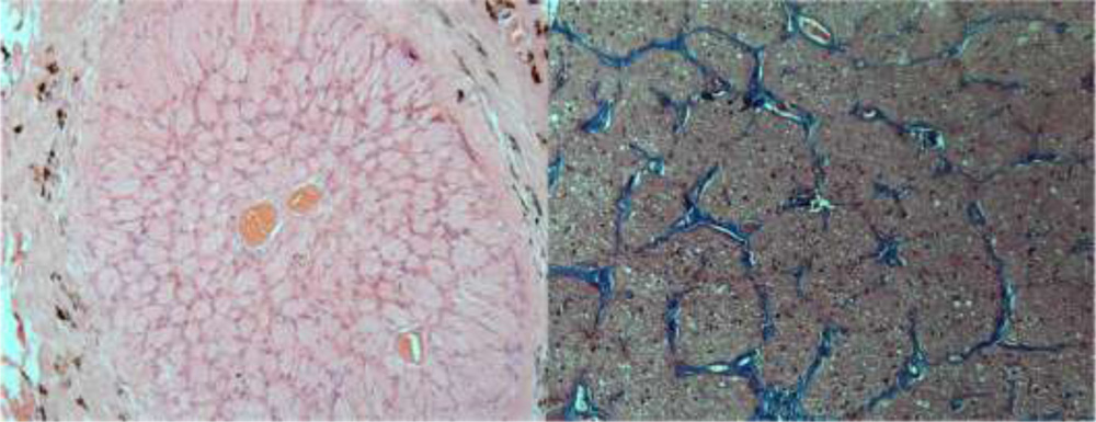
Figure 3: Optic nerve section of unstained slide, under polarizer (20X, 40X)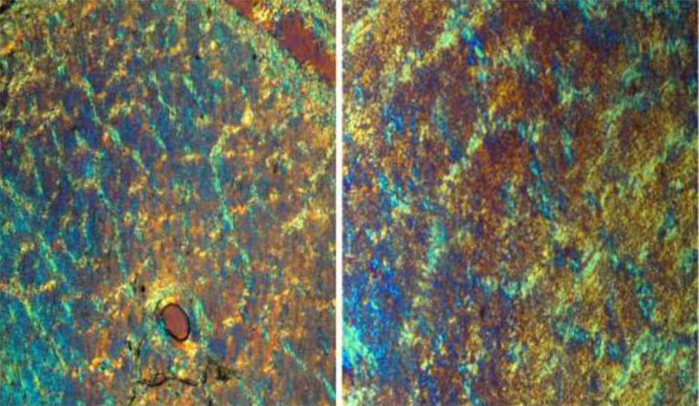
Discussion:
: Optic nerve by itself is a unique cranial nerve and till now, ‘in-vivo imaging’ to pick up the septas of the nerve and their nerve bundles seems to be remote. In future, in-vivo vital staining with magnified stereoscopic imaging can be possible to demarcate those septas and the blood vessels within them.
Anatomic characteristic uniqueness of the optic nerve at the point of slice is inimitable and it is unmatched with other optic nerve cut point visible under microscopy.
As we know from different biometric signatures of human being, the ophthalmic science has contributed two of its parts in this “identity-science”, for instance, the iris pattern and retinal vessels configuration. Our thought process was for the third biometric mark in eye in the form of optic nerve pattern. The septas of optic nerve, at the point of specific cut is so variable that makes it a unique pattern. Second point which makes it more distinct is the position of blood vessels within the septas. If we compare it with the finger printing, then the finger print and the pores within them seems homologous to the optic nerve septas and blood vessels respectively. This makes the two structures distinctive. However, finger printing is external and can be modified or reduplicated but optic nerve is internal and cannot be reached to modify it.
Limitation of the study:
Optic nerve diseases like optic atrophy or papilledema have an effect on this inimitable anatomical distribution of collagen septas and their blood vessels in the optic nerve. This might have an impact on reproducibility of these patterns during the process though they will have characteristic changes specific to the person and time . Due to lack of technology, it was difficult for us to study the effect of diseases and it’s reversibility after the disease process has subsided.
Conclusion:
Present study observed an inimitable anatomical distribution of collagen septas with their blood vessels at the point of slice of the optic nerve.
These anatomical optic nerve observations can be applicable when future in vivo imaging’s methods can be developed to demarcate those collagen septas and intra-septal blood vessels.
Selected References:
- O’ Grorman” Fingerprinting verification” Biometrics, personal identification in networked society( Anil Jain, Ruud Bole, Sharath Pankanti Eds) 1999
- R Hill “ Retinal identification” Biometrics personal identification in networked society (Anil Jain, Ruud Bole, Sharath Pankanti Eds) 1999
- Edward S Dunshire. Emerging biometric developments, identifying the missing pieces in industry
- Bernadatte Dorizzi. Biometric at the frontiers, assessing the impact on society technical impact on biometric. European communication 2005
- Trobe JD. Neurology of Vision. New York: Oxford University Press; 2001: 1-42