Dr.Sushil Bajoria, B08439,
Dr. Vijaya Jojo, Dr.
Surendra Prasad Jakhanwal
Authors:Dr. Sushil Kumar Bajoria, Dr. Vijaya Jojo, Dr. S. P. Jakhanwal
Aim:
To evaluate central corneal thickness (CCT) pre and post phacoemulsification and study its significance in relation to intraocular pressure changes.
Introduction:
Cataract surgery is amongst one of the most common surgeries performed in various parts of the world1,2. It induces changes in the corneal properties. This has led to studies being conducted in various changes that take place after cataract surgery. One of the changes that take place is increase in the central corneal thickness after cataract surgery. This increases in the immediate postoperative period and then comes down to normal levels within few weeks time3,4.
Since the corneal thickness is increased in the immediate postoperative period, intraocular pressure recordings will be erroneously high. This may lead to unnecessary treatment. 5,6. An important predictor of intraocular pressure is central corneal thickness.7,8. Thicker corneas give false high values and thinner corneas give false low values3,5.
Therefore, this study was conducted to evaluate the central corneal thickness after phacoemulsification surgery and find out the duration after which it comes back to normal.
Materials and methods:
It was a prospective study conducted from January to April 2016. A total of 73 patients were enrolled. Of these, 30 patients were males and 43 patientswere females. The age group was from 55 years to 78 years for males and 49 years to 72 years for females. The patients underwent routine eye examination preoperatively. This included visual acuity examination, intraocular pressure recording, detailed slit lamp and fundus examination after dilatation. All the patients underwent phacoemulsification with foldable intraocular lens implantation. All the surgeries were performed by the same surgeon. Peribulbar anaesthesia was given to all the patients. The central corneal thickness (CCT) was measured pre operatively, on 1stpostoperative day, at 1 week and 4 weeks interval after surgery. Central corneal thickness was measured using Ocuscan RxP machine.
The exclusion criteria were patients with ocular diseases like pre existing glaucoma, history of any prior surgery, history of trauma, patients with diabetes mellitus or any other ocular pathology or disease.
Results:
The average central corneal thickness pre operatively was 522 microns, on day 1 it was 584, at 1 week it was 537 and at 4 weeks it was 528 microns (Figure 1). The Std. Deviation was 34.5, 53.6, 36.2 and 32.1 microns preoperatively, on day 1, at 1 week and at 4 weeks respectively. Central corneal thickness gradually reduced and came back to almost pre operative level by 4 weeks. The central corneal thickness increased by 11.83 % on 1st post op day, which reduced to 2.82 % by 1st postoperative week. By the end of 4th week the CCT came back to almost the preoperative levels (1.08% increase) (Table 1).
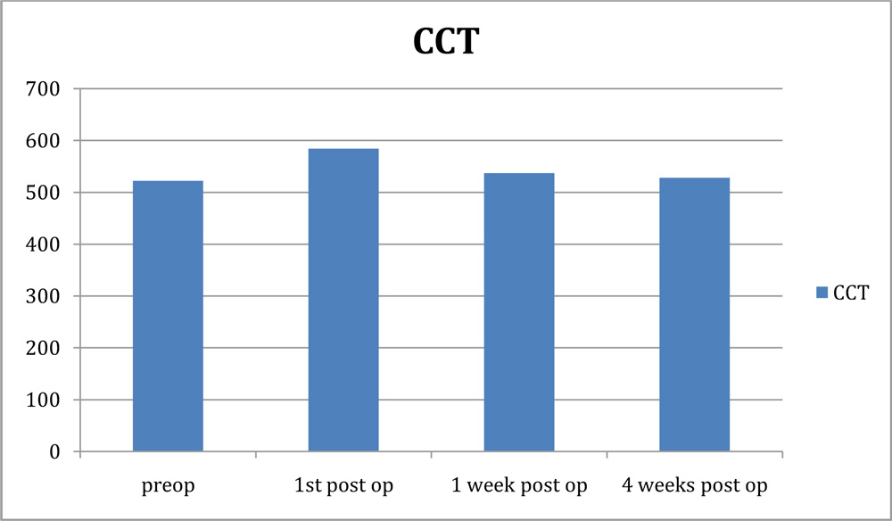
Figure1. Central corneal thickness measurement.
Table 1. showing, the mean and standard deviation of the central corneal thickness from pre-op to the 28th day
| Day | Minimum | Maximum | Mean | Std. Deviation |
| Preoperative | 458.00 | 614.00 | 522.68 | 34.53731 |
| 1st post operative day | 497.00 | 685.00 | 584.17 | 53.60339 |
| 1 wk post operative | 483.00 | 611.00 | 537.47 | 36.22211 |
| 4 wks post operative | 472.00 | 592.00 | 528.36 | 32.17406 |
Discussion:
Intraocular pressure readings are affected by central corneal thickness. If the thickness is more the readings will be falsely high and if thickness less, the readings will be falsely low.The association between intraocular pressure readings and central corneal thickness has been well documented in literature. From a central corneal thickness of 550 microns, there is approximately 1 mm Hg correction for a 25 microns deviation11.We should be aware of this relationship. The central corneal thickness has been noted by other authors to increase in the immediate postoperative period and come back to normal after 1 week following cataract surgery3.
Our study, similarly has shown that there is marked increase in the mean central corneal thickness on the first postoperative day after an uneventful phacoemulsification surgery. However, the central corneal thickness measurements come down by the first postoperative week and it is near normal by the fourth postoperative week9.This increase in central corneal thickness after cataract surgery appears to be due to corneal edema, which generally settles over the next 1 – 4 weeks9.It has been reassuringly noted by several studies that there is no significant permanent damage to the eye because of transient increase in postoperative intraocular pressure, if the eyes were healthy preoperatively10.Therefore, we also suggest that anti glaucoma treatment should not be started in the immediate postoperative period. It may be due to increased thickness of the cornea. If at all treatment is required, corrected intraocular pressure should be considered.
This study showed that the mean increase on first and seventh postoperative day was more compared to other studies for the same period (3,4). A mean increase of 3.15 μm at week 1 was reported in a study conducted by Salvi et al3. This could be because of the difference in the population studied, time taken for surgery, and the grade of cataract that were selected to be operated.
Conclusion:
Increased central corneal thickness in the immediate post operative period may give rise to false increase in intraocular pressure and unnecessary treatment.It returns to normal baseline values in almost all operated eyes in 1-month time. Therefore we should refrain from treatment as the central corneal thickness comes down rapidly especially in uncompromised eyes.
Acknowledgement:
Ms. Harshita Biswas,
M.Phil (Medical& Social Psychology).
References:
1.Taylor HR.Cataract: how much surgery do we have to do? The Br J Ophthalmology. 200; 84: 1-2.
2.Franchini, A., Frosini, S., Boddi, V. 2008. Standard coaxial phacovsmicroincisioncataract surgery: a cornealendothelium study. Int J Ophthalmol, 1(4): 344–350.
3. Salvi SM, Soong TK, Kumar BV, Hawksworth NR. Central corneal thickness changes after phacoemulsification cataract surgery. Journal of cataract and refractive surgery. 2007; 33: 1426-8.
4.Falkenberg B, Kutschan A, Wiegand W.Analysis of optical parameters after cataract surgery and implantation of foldable lens. Der Ophthalmology Zeitschrift der Deutschen Ophthalmologischen Gesellschaft 2005; 102: 587-91.
5.Bolz M, Sacu S, Drexler W, Findl O.Local corneal thickness changes after small-incision cataract surgery. Journal of cataract and refractive surgery. 2006; 32: 1667-71.
6.Recep OF, Hasiripi H, Cagil N, Sarikatipog H.Relation between corneal thickness and intraocular pressure measurement by noncontact and applanation tonometry. Journal of cataract and refractive surgery. 2001; 27: 1787-91
7.Brandt JD. The influence of corneal thickness on the diagnosis and management of glaucoma. Journal of glaucoma 2001; 10: S65-S7.
8.Chu J, Tham YC, Liao J, Zheng Y, Aung T, Wong TY, Cheng CY.Ethnic differences of intraocular pressure and central corneal thickness; the Singapore Epidemiology of Eye Diseases study. Ophthalmology 2014; 121: 2013-22.
9. TanveerA C, Muhammad H, WajihaK, Khabir A, 2015. Central Corneal Thickness Changes after Phacoemulsification. Pak J Ophthalmol, Vol. 31No. 2
10.Tranos P, Bhar G, Little B. Postoperative intraocular pressurespikes: the need to treat. Eye 2004; 18:673–679
11.Kohlhass M, Boehm AG, Spoerl E, et al. Effect of central corneal thickness, corneal curvature, and axial length on applanation tonometry. Arch Ophthalmol 2006; 124:471–476