Dr. Raju S, R09402, Dr. Hemalatha B C
Dr Raju Sampangi, Dr Hemalatha B C
Nethraspandana Eye Hospital , Bangalore
Introduction
Modern vitrectomy was introduced by Dr Robert Machemer in 1970, since then many improvements in surgical techniques and surgical instrumentation are continuously been developed. One of the most important improvements is the use of wide-angle viewing system (WAVs) during retinal surgeries. The use of WAVs gives operating retina surgeons a panoramic view of the surgical field thereby improving the safety and efficiency of the surgical procedures.
Wide angle viewing systems can be classified as contact and non-contact viewing systems. Commonly used contact lens systems are the Miniquad XL from Volk and wide field lens from Ocular instruments. Non contact wide angle viewing systems include the BIOM1 by Oculus instruments, Resight by Zeiss, EIBOS by Moller Wedel, Peyman–Wessels–Landers lens (PWL)2 and 132D Upright Vitrectomy Lens by Ocular instruments.
Each viewing system has advantages and disadvantages. PWL lens provides wide-field upright images without an inverter while the 132D lens requires the inverter. The PWL lens and 132 D lenses were originally designed in such a way that they are attached to the wrist rest of the operating table using standard clamp, straight rods, and linkage system. This is independent of the microscope. (Figure 1) This arrangement has the advantage that the focus can be adjusted with the footswitch of the microscope; however one needs to readjust the lens arrangement to maintain the view and centring i.e appropriate XYZ position. This drawback is the main reason as to why this lens system is not commonly used although it is less expensive than other noncontact lens systems.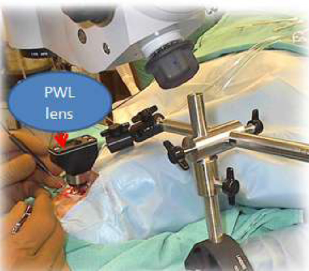
FIGURE 1
We have developed a new universal holding system for the PWL lens and 132 D to overcome the possible disadvantages of the original design.
Material and Methods
The new lens holder is attached to the microscope unlike the original attachment that is attached to the wrist rest of the operating table. It consists of 2 main parts: a vertical rod attached to the microscope and is fixed .It allows for attaching the lens holding part. The lens holder consists of a rotating bar with ring at the lower end and a system for adjusting vertical position of the rotating bar. (Figure 2)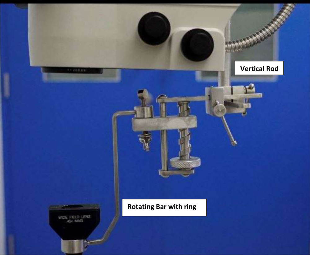
FIGURE 2
The ring at the lower end holds the PWL lens and the bar can be rotated 210 degrees, this allows for swivelling the lens into position during surgery and can be swivelled out when not in use (Figure 3). This arrangement is in turn attached to a screw based rotational system which translates into up and down movement for the rotating bar carrying the lens. This vertical movement helps to adjust the distance between the lens and the corneal surface during surgery. Closer the lens is to the cornea larger the field of view. The height can be adjusted to 1mm precision. The lens holder can be steam sterilised for surgery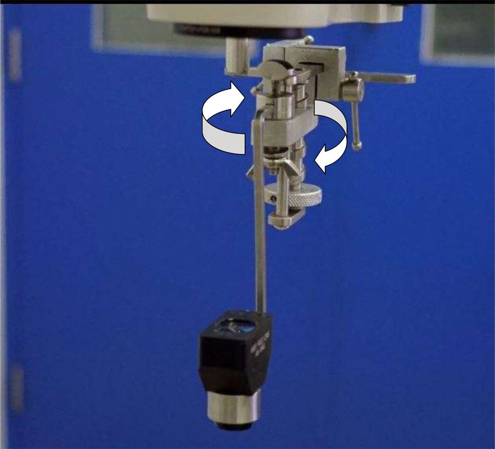
FIGURE 3
Only for the first time the surgeon needs to set up the new holding system by adjusting the position of the ring of rotating bar; once set, the lens holder is precisely held at the centre of the microscope. The PWL lens is placed in the ring of the rotating bar to get a wide angled view of the retina. The swivel design allows the surgeon to effortlessly switch between anterior segment and posterior segment during surgery just by rotating the bar. As the PWL lens comes with an inbuilt prism re-inverter there is no need for any other inverter to be attached to the microscope. Another innovation that we have done is the same lens holder with a minor modification in the rotating bar can hold the 132D lens to achieve a wide angle
viewing of the retina. This 132D lens however requires the inverter to be fixed to the microscope.(Figure 4)
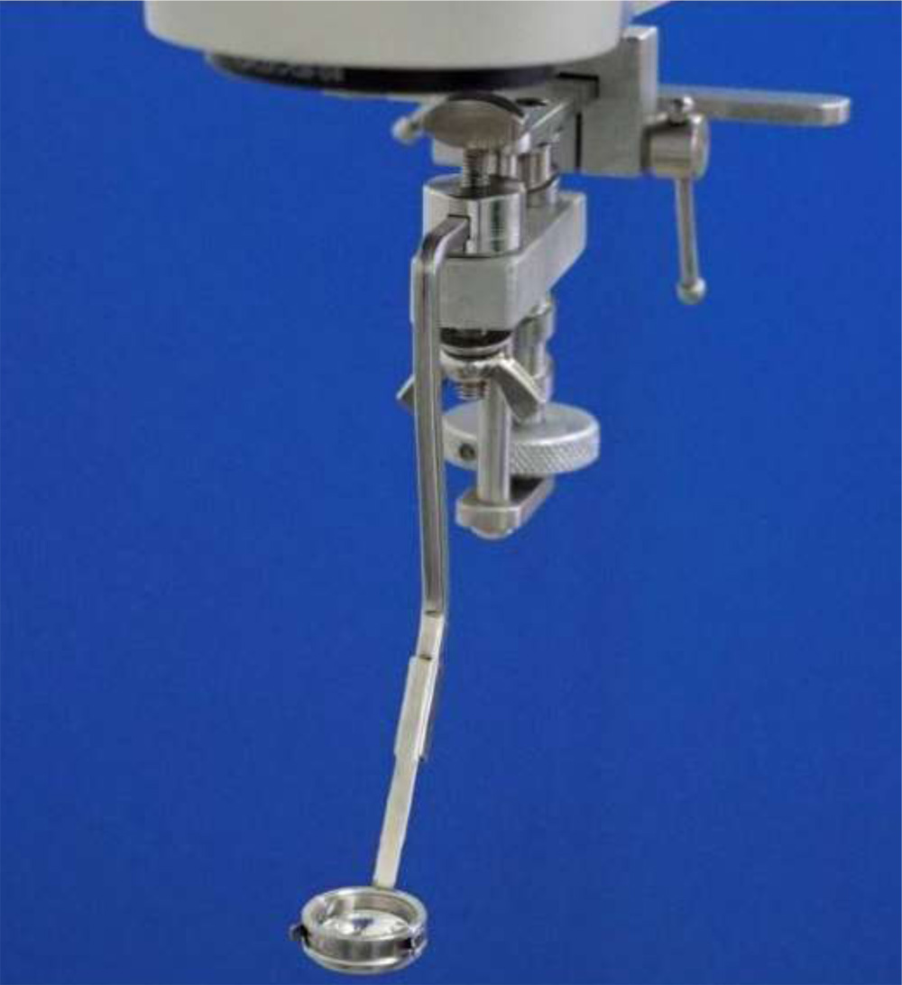 Figure 4 (Attachment with 132D lens)
The advantage of this viewing system is it can be attached to any microscope by altering the fixed part that attaches to the microscope. The view is comparable to other wide angle viewing systems for all practical purposes. Comparative wide angle retinal views with PWL lens, 132D lens and contact wide angle with Volk Miniquad Xl are show in Figures 5, 6, 7&8. One can use the microscope footswitch for focus unlike other systems where the condensing lens needs adjustment for focus. This ability to use microscope footswitch allows for very crisp retinal viewing during surgery, this also obviates the need for a condensing lens when using either PWL lens or the 132D lens.
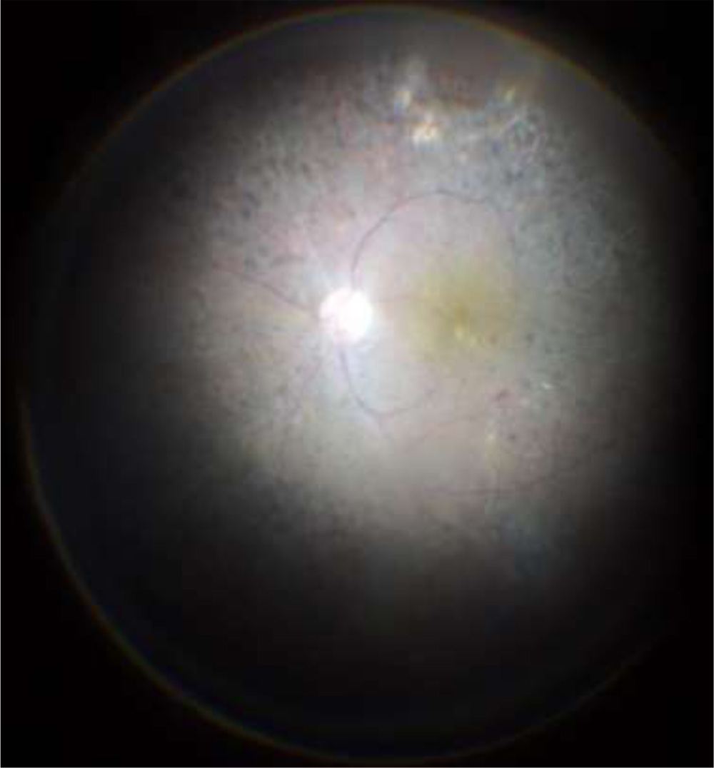 Figure 5 – View with PWL lens in fluid filled eye
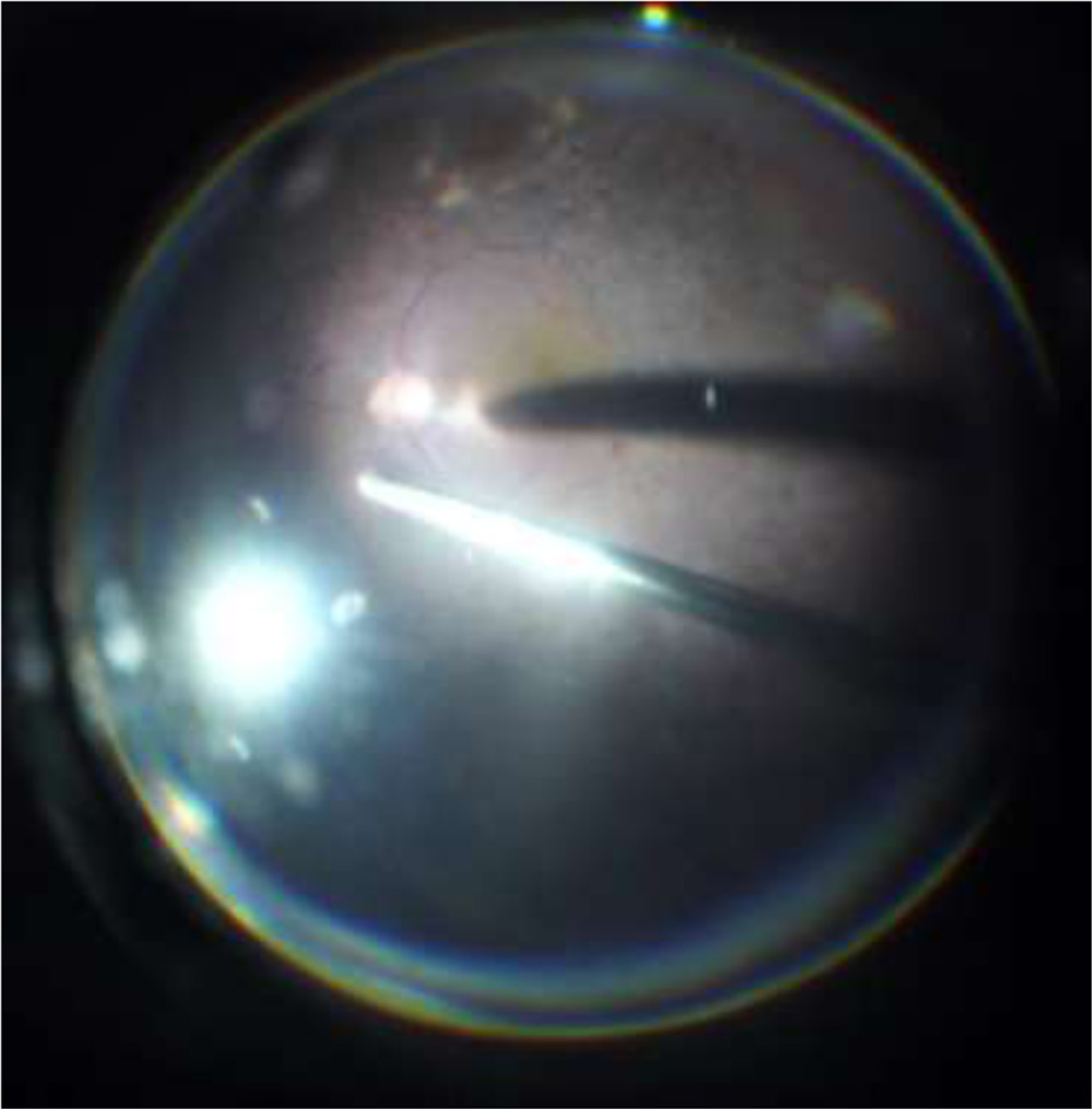 Figure 6- View with PWL lens in air filled eye
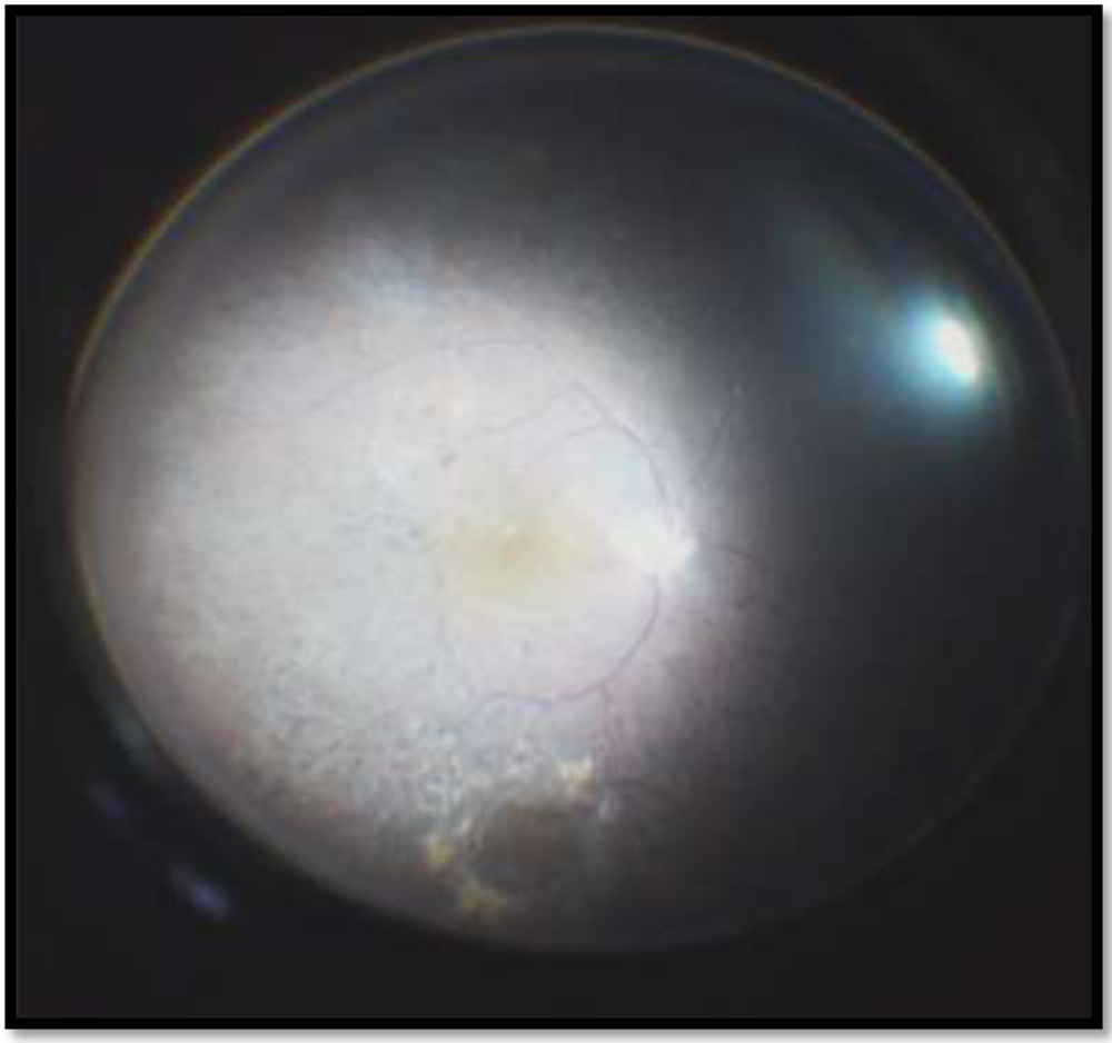 FIGURE 7: View with 132D lens in fluid filled eye
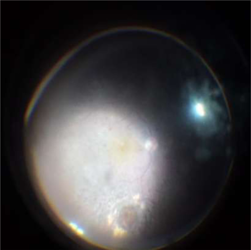
Figure 8 : View with Contact wide angle miniQuad XL for comparison
Discussion
PWL lens or 132D lens can be bought separately and can be used with our attachment. The newly developed universal lens holding system for the PWL lens/ 132D lens gives us the versatility of a non-contact wide angle viewing system and imaging capability of a contact viewing system. This combination can be a cost effective alternative to the existing viewing systems.
Additional Media.
Following youtube videos of the surgery done using this attachment can be viewed
https://www.youtube.com/watch?v=Vip8fQV0NnA
https://www.youtube.com/watch?v=KWtfGLmMhh8
https://www.youtube.com/watch?v=X4Rp1yacK0U
References.
- Spitznas M. A binocular indirect ophthalmoscope (BIOM) for non-contact wide-angle vitreous surgery. Graefes Arch Clin Exp Ophthalmol 1987;225:13–15.
- Landers MB, Peyman GA, Wessels IF, Whalen P, Morales V. A new, non-contact wide field viewing system for vitreous surgery. Am J Ophthalmol 2003;136:1191–1192.