Dr. Jyoti Gupta,G17478, Dr. Shakun Gupta, Dr. Shweta Tripathi, Dr. Alka Gupta
Chief author: Dr.Shakun Gupta¹, Presenting author:Dr.Jyoti Gupta¹,
1-Indira Gandhi Eye Hospital & Research Centre , Lucknow, UP, India
ABSTRACT
Aim:
To compare the variation in stereoscopic vertical cup disc ratio examination with 90 D lens Vs. Cirrus HD-OCT.
Material and methods: It is aretrospective study in which all cases in whom Cirrus HD-OCT was done for glaucomatous disc evaluation & examined by single experienced glaucoma specialist, in the month of January 2016 were included.It includes 94 eyes of 49 patients (4 patients were one eyed).
Difference of vertical cup disc ratio between 90 D and cirrus HD-OCT was noted, values less than 0.1 were considered as comparable while the values more than or equal to 0.1 wereconsidered as non-comparable.
Results:
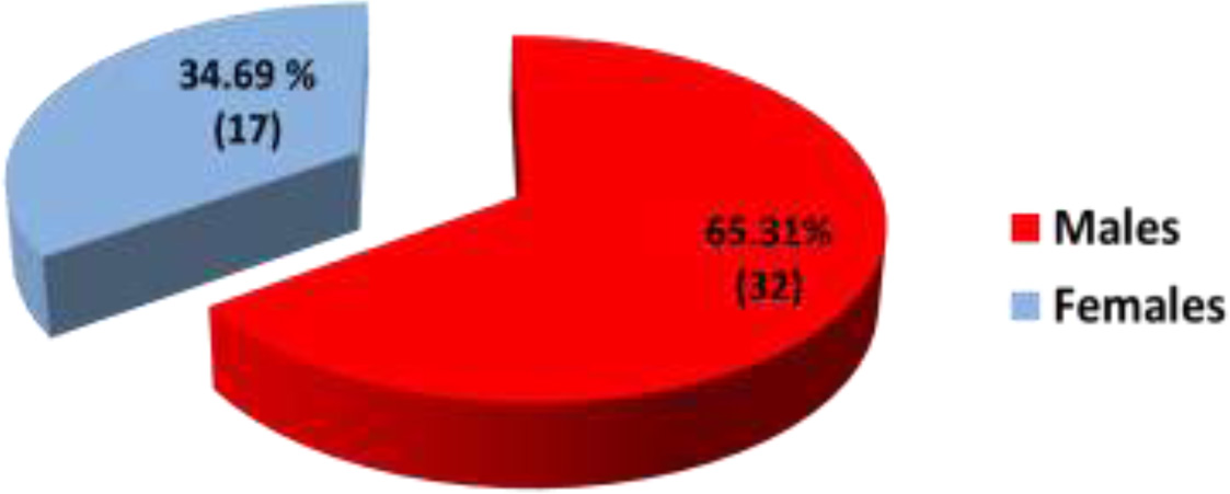
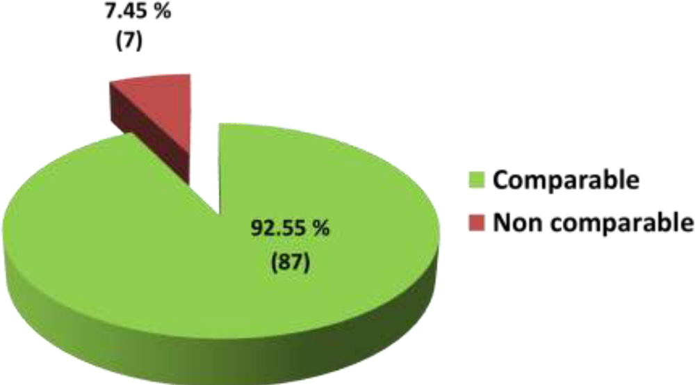
Age ofthe patients was 44.26± 17.89 years (mean ± SD). Out of 49 patients, 65.31% (32) were males and 34.69% (17) were females. Vertical Cup disc ratio was comparable in 92.55% (87) and found to be non-comparable in 7.45% (7) of eyes with Cirrus HD-OCT.
Conclusion:
Stereoscopic vertical cup disc ratio examination with 90 D lens was found to be comparable with Cirrus HD-OCT in majority of the cases.However, OCT overestimates vertical CD ratio in few cases.
Introduction:
Ophthalmoscopic estimation of the vertical cup-to-disc ratio (VCDR) of the optic nerve head is important in the management and follow-up of patients with glaucoma or glaucoma suspects. In a clinical setting, slit lamp indirect ophthalmoscopy with 90 D is frequently used for this, but it has only a moderate inter-observer agreement and relies on observer experience.1,2OCT is capable of measuring a greater number of parameters and in a more reproducible way than ophthalmoscopy.
OCT, a high-resolution non-contact imaging modality with great acceptance among ophthalmologists, is an excellent diagnostic tool for objective and quantitative measurement of ONH and RNFL parameters.3 The reproducibility of ONH measurements using the OCT (Carl Zeiss Meditec, Dublin, CA, USA) was reported to be sufficient, especially regarding the cup-to-disc ratios (CDRs).4
Cirrus HD-OCT have improved image resolution, imaging speed, and sensitivity5 compared to Stratus OCT technology. Thus, based on these technological improvements, it can be expected that Cirrus HD-OCT has superior performances in glaucoma as compared to Stratus OCT.
To the best of our knowledge, there have been no reports comparing stereoscopic vertical CDR with 90 D vs.Cirrus HD-OCT. Therefore, this study was done to compare variation in stereoscopic vertical CDR examination with 90 vs. Cirrus HD-OCT.
Aim:
To compare the variation in stereoscopic vertical cup disc ratio examination with 90 D lens Vs.Cirrus HD-OCT.
Material and methods:I
t is aretrospective study in which all cases in which Cirrus HD-OCT was done for glaucomatous disc evaluation & examined by single experienced glaucoma specialist, in the month of January 2016 were included.It includes 94 eyes of 49 patients (4 patients were one eyed).
Patients having corneal opacities, cataract, media opacities and other ocular pathology were excluded from the study. Patients whose OCT report had signal strength less than 5were also excluded from the study.
Difference of vertical cup disc ratio between 90 D and Cirrus HD-OCT was noted.
Values less than 0.1 wereconsidered as comparable while the values more than or equal to 0.1 wereconsidered as non-comparable.6
Results:
Sex distribution: Out of 49 patients, 65.31% (32) were males and 34.69% (17) were females.
Age distribution: Age of the patients was 44.26± 17.89years (mean ± SD).
| Years | Percentage (Numbers) |
| 1 – 20 | 12.24 % (6) |
| 21 – 40 | 26.53 % (13) |
| 41 – 60 | 38.77 % (19) |
| 61 – 80 | 22.44 % (11) |

Vertical CDRcomparison: Vertical cup disc ratio were comparable in92.55% (87) and found to be non-comparable in 7.45 % (7) of eyeswith Cirrus HD-OCT.
Discussion:
No study was found comparing stereoscopic vertical CDR with 90D vs. Cirrus HD-OCT. However, few studies are available which has done comparison of CDR estimated by OCT vs. other methods.
Dr.Maragatham et al compared the difference of cup disc ratio between direct ophthalmoscope and OCT with approximately same method as of ours and found OCT overestimates the CDR in 61% of the glaucoma suspects and 52% of established glaucoma respectively. Thus, clinical examination is more important. So, OCT can be used as an adjunct in diagnosing glaucoma.6
Arnalich-Montiel et al7 studied cup-to-disc ratio agreement between slit-lamp indirect ophthalmoscopic estimation and stratus optical coherence tomography measurement and found OCT shows higher values than the specialists; the greatest differences occurred when assessing small CDRs and the differences diminished as the cupping increased. These two methods of measurement are not interchangeable, and the difference must be considered, especially in discs with smaller CDRs.
Prof. [Dr.] MeenakshiDhar et al8 compared the results of optic disc analysis using optical coherence tomography (stratus OCT model 3000), fundus photography and stereoscopic biomicroscopy and found 50% cases showed good correlation between all the 3 methods. Optical coherence tomography showed a higher valueindicating the requirement to do optical coherencetomography in all patients to detect the actual cup disc ratio which will help us to detect glaucoma cases earlier and to treat them, well before axonal loss occurs.
Difference between our study and above studies may be because Dr.Maragatham et al6 compared two dimensional CDR by direct ophthalmoscope to stratus OCT while we compared three dimensional VCDR with 90D to Cirrus HD-OCT & with that of Arnalich-Montiel et al7 and MeenakshiDhar et al8may be because of different methods & different OCT used as Cirrus HD-OCT has more almost twice image resolution, more imaging speed, and sensitivity as compared to stratus OCT.
Jason Dobson et al9 did comparison of vertical cup-to-disc ratio using fundus photography vs. Cirrus SD-OCT & found excellent correlation (0.800) between clinician VCDR grading using fundus photography vs. Cirrus SD-OCT.
Difference between our & their study is that they did vertical CDR grading using fundus photography while we did stereoscopic slit lamp indirect ophthalmoscopy with 90D and both of us compared VCDR with VCDR of Cirrus HD-OCT.
Our study also showsexcellent strength of agreement (0.925)between stereoscopic vertical CDR with 90D vs. Cirrus HD-OCT according to Fiess’ Kappa Benchmark Scale.10
Limitation of study: It has small number of samples. Further studies are required with large number of samples.
Conclusion: Stereoscopic vertical CD ratio examination with 90 D lens was found to be comparable with Cirrus HD-OCT in majority of the cases. However, OCT overestimates vertical CD ratio in few cases.
References:
1)Lichter PR. Variability of expert observers in evaluating the optic disc. Trans Am OphthalmolSoc 1976; 74: 532–572.
2)Tielsch JM, Katz J, Quigley HA, Miller NR, Sommer A. Intraobserver and interobserver agreement in measurements of optic disc characteristics. Ophthalmology 1988; 95: 350–356.
3)Schuman JS, Hee MR, Arya AV, Pedut-Kloizman T, Puliafito CA, Fujimoto JG et al. Optical coherence tomography: a new tool for glaucoma diagnosis. CurrOpinOphthalmol 1995; 6: 89–95.
4)Medeiros FA, Zangwill LM, Bowd C. Evaluation of retinal nerve fiber layer, optic nerve head, and macular thickness measurements for glaucoma detection using optical coherence tomography. Am J Ophthalmol 2005; 139: 44– 55
5)Chen TC Cense B Pierce MC. Spectral domain optical coherence tomography: ultra-high speed, ultra-high resolution ophthalmic imaging. Arch Ophthalmol. 2005; 123: 1715–1720.
6)Comparison of Cup Disc Ratio Ophthalmoscopically and Optical Coherence Tomography wiseDr. Maragatham K., Dr.Vijay Krishnan B., 70th AIOC proceedings, Cochin 2012
7)Cup-to-disc ratio: agreement between slit-lamp indirect ophthalmoscopic estimation and stratus optical coherence tomography measurementArnalich-Montiel F1, Muñoz-Negrete FJ, Rebolleda G, Sales-Sanz M, Clinical Study,Eye (2007) 21, 1041–1049; doi:10.1038/sj.eye.6702391; published online 5 May 2006
8)Comparison Of Results Of Optic Disc Analysis Using Stereoscopic Biomicroscopy, Stereo Fundus Photography and Optical Coherence Tomography Prof. [Dr.] MeenakshiDhar, Dr. InduJayachandran, Dr. BijuRaju, Ms. Deepa PA
9)Comparison of vertical cup-to-disc ratio and optic nerve head size usingfundus photography vs. Cirrus SD-OCT, Jason Dobson, Derek Wills, Leland Carr, ODNortheastern State University Oklahoma College of Optometry
10) Benchmarking Inter-Rater Reliability Coefficients, chapter 6,page 125
Authors have no financial interest.
Correspondence to: Dr. Shakun Gupta
E-mail id – drshakunsnc@rediffmail.com