Dr. Shail, V13188,
Dr. Viraj Abhayakumar Vasavada, Dr. Vasavada
Abhaykumar Raghukant, Dr. Samaresh
Abstract:
Outcomes afterPhaco versus FemtoLaser-Assisted Phacoemulsification in Shallow Anterior Chambers
Authors:
Shail, Viraj, ARV, Samaresh
Synopsis:
Prospective,randomized clinical trial to compare intraop performance and postop outcomes in eyes with shallow anterior chamber(ACD<2.5mm) undergoing femto laser assisted cataract surgery (FLACS)(n=91) vsphaco(n=91)
.Outcome measure: Corneal thickness(CCT) on day 1,week 1, 1 month,Cornealclarity,AC cell-flare,Descemet folds on day 1,week 1,1 month,Change in Endothelial cell density at 3 months,Vision on day1,1 month.
Results: Day 1,mean CCT in Group 1 and 2 was 565+35u and 589+28u (P=0.03). The % change in CCT at day 1,Group 1 and 2 was 11% and 19% (P=0.04).There was no significant difference in the mean CCT or the percentage change between groups at 1 week and 1 month.Eyes with >Grade 2ACcells was higher in Group 2 compared to Group 1 at day 1(30% vs23%) and 1 week(27% vs 20%), there was no difference at 1 month.Change in endothelial cell density was not significant between two groups.FLACSleads to lesser AC inflammation and lesser increase in CCT on day 1,in shallow AC
A shallow anterior chamber (<2.5mm) is a challenge during cataract surgery, because it reduces the already tight confines of the anterior segment of the eye. Few authors indicated shallow anterior chamber as a factor responsible for increased rates of peri-operative complications in these patients1,2,4. Day and associates have shown a strong relation between shallow anterior chamber and peri-operative complications. Also such patients are likely to have a longer recovery time post-operatively. Further in these eyes a narrow anterior chamber angle is thought to be important because a long standing narrow angle or appositional closure may predispose to peripheral anterior synechiae formation, which may lead to uncontrollable rise in intra-ocular pressure (IOP).
Femtosecond Laser Assisted Cataract Surgery (FLACS) and its advantages in routine as well as difficult cataract surgery are coming up more and more4. FLACS has the advantage of performing an appropriate sized and centered capsulorhexis as well lens fragmentation. This reduces the mechanical manipulations that need to be done inside the anterior chamber as some of the important steps are already pre-performed. Often , manipulation with the cystitome in a shallow chamber can lead to small or large decemts detachment sub-incisionally.
So we decided to compare the intra-operative performance and post- operative outcomes in eyes undergoing femtosecond laser assisted cataract surgery versus eyes undergoing phacoemulsification in eyes having shallow anterior chamber
Materials and Methods:
This was a Prospective Randomised clinical trial. All patients having age related cataract and a shallow anterior chamber (<2.5mm) were included. All patients having any other ocular co-morbidity were excluded from the study. A single surgeon (ARV), standardized surgical technique was used in all cases. The Femtosecond platform that was used was LenSx (Alcon Laboratories, USA) and phacoemulsification was performed using the Centurion Vision System (Alcon Laboratories, USA).
Total 182 patients were included in the study. Group 1 were patients who were randomised to undergo FLACS (n=91) and Group 2 were patients who were randomised to undergo routine phacoemulsification. In Group 1, capsulorhexis and Lens fragmentation (chop pattern) were performed in all patients with the LenSx. Sideport and Main corneal incisions were made using knives in both groups.
Statistical Analysis was done using paired t test and SPSS software..
Outcome Measures:
Primary Outcome Measures : Central Corneal Thickness (CCT) was measured pre-operatively, and then post-op Day 1, 1 week, 1 month and 3 months. CCT was used as a primary outcome measure as it is one of the most sensitive indicators of corneal edema, which is an indirect evidence of endothelial trauma during surgery. CCT was measured in all cases using the Anterior Segment OCT (Carl Zeiss Meditec, USA).
Secondary Outcome Measures: Anterior chamber cells and flare (hogan’s criteria), Corneal Clarity (slit lamp grading), Corneal endothelial cell count. All of these parameters were noted at post-op Day1, 1 week, 1 month and 3 months post-operatively.
Results:
The mean age in Group 1 was 67.21 +/- 11.11 and mean age in Group 2 was 63.70 +/- 11.81. There was no statistical difference between the 2 groups.
| Group 1 | Group 2 | P value | |
| Pre-op | 2.38 +/- 0.13 | 2.27 +/- 0.15 | 0.60 |
| Post-op | 3.46 +/- 0.48 | 3.48 +/- 0.39 | 0.56 |
Table 1: Anterior chamber depth
The CCT was increased in both groups post-operatively compared from pre-operatively. However, CCT increase was less in Group 1 on day 1 and 1 week post-operatively compared to Group 2. This difference was statistically significant. At 1 month, there was no difference in CCT between the 2 groups.
| Pre-op | POD-1 | POD 1 week | POD 1 month | |
| Group 1 | 531.2 +/- 42.6 | 540.40 +/-49.40 | 535.5 +/- 44.3 | 532.3 +/- 40.9 |
| Group 2 | 528.6 +/- 39.2 | 556 +/- 12.5
|
551+/- 40.8
|
530.1 +/- 39.6
|
| P <0.03 | P<0.05 | P=0.66
|
Table 2: CCT Comparison between 2 groups at different time points
Corneal Clarity was graded on the slit lamp by a masked observer post-operatively. The grading system used has been validated in literature (1- Clear cornea, 2- DM folds, 3- Focal stromal edems, 4- Diffuse stromal edema). There was a statistically significant difference between the 2 groups, group 1 had significantly clearer corneas on Day 1 and Day 7. There as no difference between the 2 groups at 1 month post-operatively.
Corneal Clarity on Day 1
Significantly Clearer Corneas in Femtosecond Group on the 1st Day and at 1week
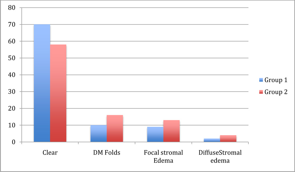
No difference at 1 month
Anterior Segment Inflamation was statistically significantly less in Group 1 at Day 1. This differene was not maintained at 1 week and 1 month post-operatively.
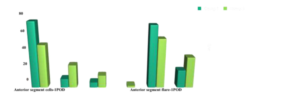
Anterior Segment Inflammation significantly lower at day 1 in FLACS group
A specular microscopy was performed in all eyes pre-operatively, post-operatively. There was a trend towards less endothelial cell loss in Group 1, however this difference was not statistically significant at 3 months post-operatively.
| ECD | CV | 6A | ||||
| Pre – op | Post op 3 months | Pre – op | Post op 3 months | Pre – op | Post op 3 months | |
| Group 1 | 2351.8±405.4
|
2157.8±392.7
|
32.7±6.7
|
30.6±6.6
|
53.4±12.3
|
51.4±10.2
|
| Group 2 | 2493.5±394.8
|
2246.6±570.3
|
30.7±6.3
|
30.2±6.5
|
54.1±10.4
|
54.0±9.1
|
Table 3: Corneal Endothelial cell count between the 2 groups
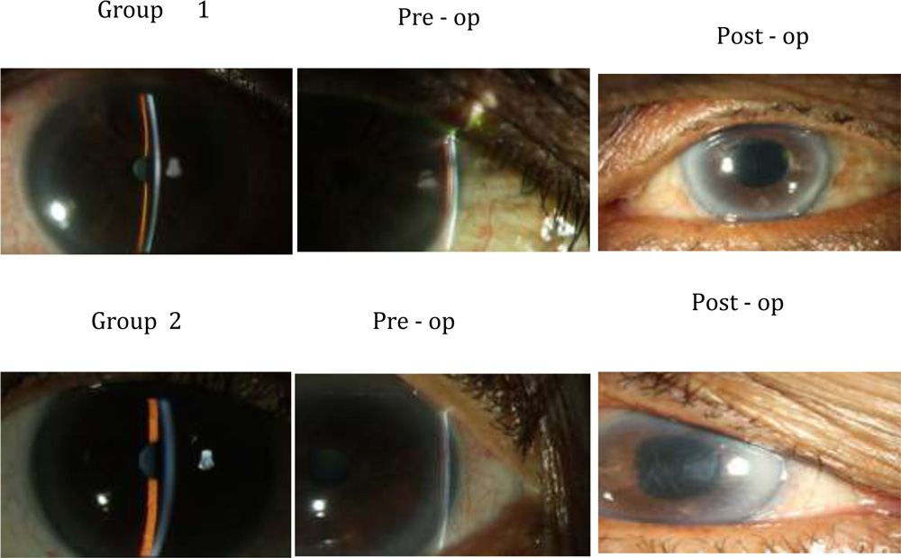
Following are some of the representative clinical photographs :
Discussion:
Cataract surgery in today’s time has reached a stage where an excellent clinical outcome on post-op day 1 is the norm. Patients with shallow anterior chamber, due to a difficult surgical environment, tend to have more corneal edema and chances of a longer post-operative recovery time. Therfore, these patients need to be counseledextensively in the pre-operative period about the possible post-operative events.
FLACS leads to lesser anterior segment inflammation, lesser corneal edema and significantly clearer corneas compared to conventional phacoemulsification. This may lead to a smoother post-operative courseand faster visual recovery. Also, with 2 important steps of surgery already been performed, it reduces intra-operative manipulation, reduces the amount of phaco energy and improves the surgical performance. Less chair time may be needed to counsel these patients pre-operativley for sub-optimal visual outcome in the post-operative period. There is a lot of debate about the utility of FLACS in routine cataract surgery, particularly in the hands of experienced cataract surgeons. However, the importance of FLACS in difficult situations like posterior polar, white cataract, shallow anterior chamber cannot be ignored. The only limiting factor today for more widespread adoption of FLACS is the prohibitively high cost of acquiring and maintaining the technology.
References:
1)Endothelial Cell Loss after Phacoemulsification according to Different Anterior Chamber Depths.Hwang HB1, Lyu B1, Yim HB1, Lee NY1. J Ophthalmol. 2015;2015:210716
2) Analysis of refractive status after cataract surgery in age-related cataract patients with shallow anterior chamber. Zhonghua Yan KeZaZhi. 2014 Feb;50(2):84-8.
3)Femtosecond laser cataract surgery: challenging cases.Martin AI1, Hodge C, Lawless M, Roberts T, Hughes P, Sutton G.CurrOpinOphthalmol. 2014 Jan;25(1):71-80
4)Endothelial Cell Loss after Phacoemulsification according to Different Anterior Chamber Depths.Hwang HB1, Lyu B1, Yim HB1, Lee NY1. J Ophthalmol. 2015;2015:210716