Dr. Jyoti Matalia, M10661, Dr.Arkasubhra Ghosh, Dr. Vimal Krishna Rajput, Dr.Shetty Bhujang K
Purpose:
With a prevalence of 15 per 10,000 children, pediatric cataract is a major cause of childhood blindness worldwide. Although various mutations have been identified in familial studies, the mechanistic basis of cataract development remains poorly understood. There have been no studies related to gene expression in pediatric cataract. We evaluate the expression of pro-fibrotic factors, structural proteins and developmental transcription factors in phenotypically & etiologically distinct forms of pediatric cataract.
Methods:
Lens material was sampled during routine pediatric cataract surgery with prior approval of the Institutional Ethics Committee and written informed consent. Lens material was sampled during cataract surgery (n=53), from which mRNA was extracted & converted to complementary-DNA. Expression of 11 genes was analyzed with the help of q-PCR.
We classified pediatric cataract and established 8 distinct groups of pediatric cataract as follows:
- Prenatal infectious cataract secondary to;
- cytomegalovirus,
- rubella
- combined cytomegalovirus with rubella
- Prenatal non-infectious cataract
- Posterior capsular anomalies,
- Postnatal developmental cataract,
- Traumatic cataract,
- Secondary cataract
The children who had cataract since birth were included in prenatal group and those who developed it later in life were categorized as developmental cataract. The congenital group was further subdivided into infectious (TORCH positive) and non-infectious (TORCH negative) groups, based on the TORCH profile of the child. Those in the secondary group had developed cataract secondary to radiation therapy/long term use of steroids/ high myopia. The patients who had only posterior capsular pathology (for example, posterior lenticonus) where the rest of the lens was clear were kept in a separate group. All the traumatic cataract cases had history of corneal tear repair and had developed cataract following tear of the anterior capsule. Children with subluxated lens who underwent clear lens aspiration served as control (n=6).
Selection of genes was on basis of a literature search. Genes that were reported of being associated with adult cataract or had known mutations that were known to cause cataract were selected: Expression of the following 11 genes was quantified to look for any association with pediatric cataract.
- Lens structural genes:
- Aquaporin 0 (Aqp-0),
- Heat Shock protein 4 (HSPA4),
- Crystallin gamma C (CRYGC)
- Transcriptional genes:
- Musculoaponeurotic fibrosarcoma oncogene (MAF),
- Tumor domain containing 7 (TDRD 7),
- Forkhead box (FOXE3),
- Pituitary homeobox 3 (PITX-3),
- Profibrotic genes:
- Transforming growth factor beta (TGF-β),
- Alpha Smooth muscle actin (α-SMA)
- Bone Morphogenetic Protein 7 (BMP-7)
Results:
We describe a new classification of pediatric cataract into 8 groups and compared their gene expression. A specific trend of gene expression was noted in different groups of cataract
(Figure 1) when compared to controls.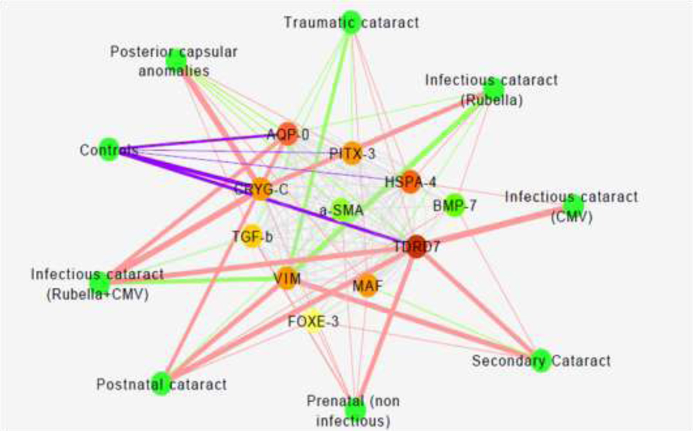
Figure 1: Co-expression network of the 11 genes based on Pearson correlation values in each disease and network of significantly highly co-expressed genes (only genes with Pearson correlation >0.95 and p-value<0.05). The red color represents down-regulation (decreased expression), green color shows up-regulation (increased expression), and purple color shows the expression in control subjects. The thickness of the lines represents the value of correlation or expression (fold change).
The prenatal cataracts were likely derived from the problems in structural genes (correlated with p=0.02, p=0.001 and p=0.003) that could be genetic in nature. The postnatal cataract showed the evidence of TGF-β driven profibrotic mechanism (correlated with p=0.002 and p=0.001) that leads to cataract formation. The infectious cataract show different profile of a high transcriptional activity with the cytomegalovirus having the highest expression of all factors (correlated with p=0.003, p= 0.006 and p=0.04) as compared to rubella (correlated with p=0.001, p=0.02, p=0.04).
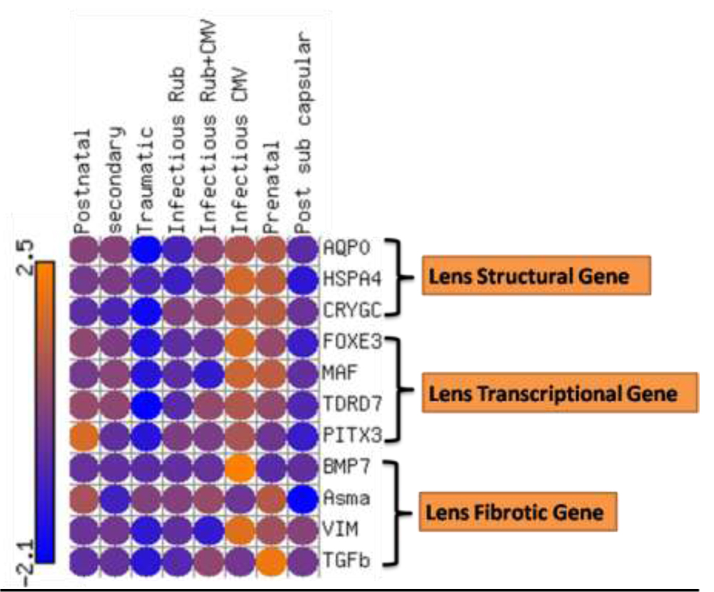
Figure: 2. This is the bird eye view of the gene expression profile that shows that mechanisms are different with different types of cataract with up-regulation in infectious CMV while it is down-regulated with posterior subcapsular and traumatic cataract.
Conclusions:
This is the first study of gene expression in pediatric cataracts that illustrates the differences in biological pathways leading to each type of cataract with correlation. We also provide a pediatric cataract classification and its correlation with the molecular expression pattern for the first time. It may eventually find its clinical application in early detection and management of pediatric cataract.